info@aethelcare.com +91 97959 65791 24/7 International Support
NEURO

Neuro surgery in India

AethelCare Provide Top Specialist Doctors And Best Hospitals For Your Medical HealthCare.
OVERVIEW

Neuro surgery? Brain & spine surgeries with precision.

Advanced neuro-navigation, minimally invasive spine surgery, micro-vascular brain tumor resection, stroke intervention, and comprehensive neurological rehabilitation.
World-Class InfrastructureNABH/JCI accredited centers with global standards
Up to 80% SavingsDramatically lower cost than USA, UK, or Australia
Expert SpecialistsInternationally trained & board-certified specialists
Priority CareImmediate scheduler booking with zero waiting list
Specialist Doctor in India
500+ Top Specialists 20+ JCI & NABH Accredited Hospitals
SPECIALIZED CONDITIONS & PROCEDURES

Explore Neuro surgery Sub Categories

Explore dedicated sub-specialties with expert doctors, accredited hospital centers, and tailored treatment plans in India.

36 Sub Specialties Get Treatment Opinion
Craniotomy for Brain Tumor Removal
Neuro surgery

Craniotomy for Brain Tumor Removal

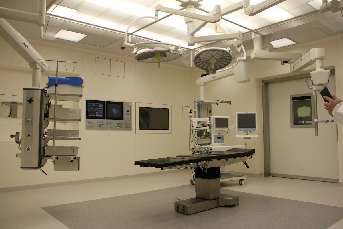
What is Craniotomy for Brain Tumor Removal? A craniotomy for brain tumor removal is a major neurosurgical procedure where a temporary bone window is created in the skull to access the intracranial vault and safely excise benign or malignant tumors. Surgeons utilize state-of-the-art neuronavigation systems (GPS for the brain), intraoperative ultrasound, and high-magnification surgical microscopes to differentiate tumor boundaries from healthy eloquent brain tissue. Indications & Common Reasons for Surgery This advanced neurosurgical intervention is recommended for patients diagnosed with: High-grade and low-grade gliomas causing neurological deficits or raised intracranial pressure. Meningiomas compressing cerebral cortex or cranial nerve pathways. Single or accessible multiple brain metastases from lung, breast, or melanoma primaries. Intracranial tumors causing intractable secondary epileptic seizures. Surgical Techniques & Precision Modalities Includes stereotactic image-guided craniotomy, awake craniotomy with intraoperative cortical stimulation for speech and motor mapping, and keyhole minimally invasive craniotomies. The procedure is performed in advanced modular operating suites equipped with 3D neuronavigation, high-definition intraoperative microscopes, neuro-endoscopy, and real-time electrophysiological monitoring to preserve vital neurological functions. Postoperative Care & Recovery Timeline Patients spend 24 hours in neuro-ICU for continuous monitoring, with typical hospital stay of 3 to 5 days and resumption of non-strenuous activities within 3 to 6 weeks. Patients benefit from specialized neuro-intensive care, personalized neuro-rehabilitation, and multidisciplinary follow-up to support optimal long-term functional outcomes. Key Clinical Benefits Targeted, maximal safe anatomical correction and pathology excision. Protection of eloquent brain areas and essential cranial nerve pathways. Significant alleviation of neurological symptoms and prevention of secondary brain injury. Accelerated recovery supported by modern microsurgical and minimally invasive techniques.

Brain Tumor Surgery
Neuro surgery

Brain Tumor Surgery

What is Brain Tumor Surgery? Brain tumor surgery encompasses comprehensive neurosurgical interventions aimed at achieving maximal safe resection of primary and secondary neoplasms of the central nervous system. The goal is to debulk tumor mass, relieve mass effect, obtain definitive histopathological and molecular diagnosis, and improve survival while preserving neurological function. Indications & Common Reasons for Surgery This advanced neurosurgical intervention is recommended for patients diagnosed with: Primary brain neoplasms including glioblastoma multiforme, astrocytomas, and ependymomas. Benign extra-axial neoplasms such as schwannomas, craniopharyngiomas, and dermoid cysts. Metastatic brain lesions causing localized cerebral edema and focal deficits. Tumors producing progressive visual loss or cognitive deterioration. Surgical Techniques & Precision Modalities Utilizes Cavitron Ultrasonic Surgical Aspirator (CUSA), intraoperative magnetic resonance imaging (iMRI), and 5-ALA (fluorescence-guided tumor visualization). The procedure is performed in advanced modular operating suites equipped with 3D neuronavigation, high-definition intraoperative microscopes, neuro-endoscopy, and real-time electrophysiological monitoring to preserve vital neurological functions. Postoperative Care & Recovery Timeline Hospital stay of 3 to 7 days followed by multidisciplinary tumor board review for adjuvant radiotherapy, chemotherapy, or targeted therapies. Patients benefit from specialized neuro-intensive care, personalized neuro-rehabilitation, and multidisciplinary follow-up to support optimal long-term functional outcomes. Key Clinical Benefits Targeted, maximal safe anatomical correction and pathology excision. Protection of eloquent brain areas and essential cranial nerve pathways. Significant alleviation of neurological symptoms and prevention of secondary brain injury. Accelerated recovery supported by modern microsurgical and minimally invasive techniques.

Meningioma Surgery
Neuro surgery

Meningioma Surgery

What is Meningioma Surgery? Meningioma surgery is the microsurgical excision of slow-growing tumors arising from the arachnoid cap cells of the meninges surrounding the brain and spinal cord. Though the majority are histologically benign (WHO Grade I), their expansion creates substantial pressure on adjacent neurovascular structures. Indications & Common Reasons for Surgery This advanced neurosurgical intervention is recommended for patients diagnosed with: Symptomatic meningiomas located along the cerebral convexity, parasagittal sinus, or falx cerebri. Skull base meningiomas (olfactory groove, sphenoid wing, petroclival) causing cranial neuropathies. Rapidly growing or atypical (WHO Grade II/III) meningiomas. Tumor-associated vasogenic edema provoking persistent severe headaches or seizures. Surgical Techniques & Precision Modalities Aims for complete Simpson Grade I resection (removal of tumor, dural attachment, and abnormal bone) using microdissection and bipolar coagulation. The procedure is performed in advanced modular operating suites equipped with 3D neuronavigation, high-definition intraoperative microscopes, neuro-endoscopy, and real-time electrophysiological monitoring to preserve vital neurological functions. Postoperative Care & Recovery Timeline Hospitalization spans 3 to 5 days, with complete long-term remission in over 85–90% of completely excised benign meningiomas. Patients benefit from specialized neuro-intensive care, personalized neuro-rehabilitation, and multidisciplinary follow-up to support optimal long-term functional outcomes. Key Clinical Benefits Targeted, maximal safe anatomical correction and pathology excision. Protection of eloquent brain areas and essential cranial nerve pathways. Significant alleviation of neurological symptoms and prevention of secondary brain injury. Accelerated recovery supported by modern microsurgical and minimally invasive techniques.

Glioma Surgery
Neuro surgery

Glioma Surgery

What is Glioma Surgery? Glioma surgery is a specialized neuro-oncological operation for resecting tumors that originate from glial support cells (astrocytes, oligodendrocytes, ependymocytes) within the cerebral parenchyma. Because gliomas infiltrate surrounding brain tissue, surgery requires meticulous microdissection to maximize tumor removal while protecting functional neural networks. Indications & Common Reasons for Surgery This advanced neurosurgical intervention is recommended for patients diagnosed with: Diffuse low-grade gliomas (WHO Grade II) prone to malignant transformation. High-grade anaplastic astrocytomas (Grade III) and glioblastoma multiforme (Grade IV). Oligodendrogliomas with IDH mutation and 1p/19q codeletion. Gliomas presenting with drug-resistant focal seizures or progressive hemiparesis. Surgical Techniques & Precision Modalities Combines awake craniotomy, subcortical white-matter tractography (DTI), functional MRI mapping, and real-time intraoperative neuro-navigation. The procedure is performed in advanced modular operating suites equipped with 3D neuronavigation, high-definition intraoperative microscopes, neuro-endoscopy, and real-time electrophysiological monitoring to preserve vital neurological functions. Postoperative Care & Recovery Timeline Inpatient monitoring for 3 to 5 days followed by personalized neuro-rehabilitation and adjuvant chemoradiation protocols. Patients benefit from specialized neuro-intensive care, personalized neuro-rehabilitation, and multidisciplinary follow-up to support optimal long-term functional outcomes. Key Clinical Benefits Targeted, maximal safe anatomical correction and pathology excision. Protection of eloquent brain areas and essential cranial nerve pathways. Significant alleviation of neurological symptoms and prevention of secondary brain injury. Accelerated recovery supported by modern microsurgical and minimally invasive techniques.

Endoscopic Brain Surgery
Neuro surgery

Endoscopic Brain Surgery

What is Endoscopic Brain Surgery? Endoscopic brain surgery is a cutting-edge minimally invasive technique that accesses intracranial pathologies through small burr holes or natural anatomical corridors using rigid high-definition endoscopes. Angled optics allow neurosurgeons to see around anatomical corners without extensive brain retraction. Indications & Common Reasons for Surgery This advanced neurosurgical intervention is recommended for patients diagnosed with: Colloid cysts of the third ventricle causing intermittent acute hydrocephalus. Intraventricular arachnoid cysts and cystic lesions. Neurocysticercosis and intraventricular parasitic cysts. Diagnostic biopsy of deep-seated periventricular and thalamic lesions. Surgical Techniques & Precision Modalities Transventricular neuroendoscopy utilizing 0-degree and 30-degree scopes, micro-balloons, and endo-bipolar coagulators through small 10mm burr holes. The procedure is performed in advanced modular operating suites equipped with 3D neuronavigation, high-definition intraoperative microscopes, neuro-endoscopy, and real-time electrophysiological monitoring to preserve vital neurological functions. Postoperative Care & Recovery Timeline Minimal postoperative pain, day-care or 24-48 hour hospital stay, and rapid resumption of everyday life within 7 to 10 days. Patients benefit from specialized neuro-intensive care, personalized neuro-rehabilitation, and multidisciplinary follow-up to support optimal long-term functional outcomes. Key Clinical Benefits Targeted, maximal safe anatomical correction and pathology excision. Protection of eloquent brain areas and essential cranial nerve pathways. Significant alleviation of neurological symptoms and prevention of secondary brain injury. Accelerated recovery supported by modern microsurgical and minimally invasive techniques.

Endoscopic Brain Tumor Surgery
Neuro surgery

Endoscopic Brain Tumor Surgery

What is Endoscopic Brain Tumor Surgery? Endoscopic brain tumor surgery utilizes specialized high-definition endoscopes to resect intraventricular, pineal, and deep cranial tumors through minimally invasive keyhole corridors, avoiding the morbidities associated with large open craniotomies. Indications & Common Reasons for Surgery This advanced neurosurgical intervention is recommended for patients diagnosed with: Intraventricular ependymomas and subependymal giant cell astrocytomas (SEGA). Pineal region tumors requiring tissue biopsy and concurrent CSF diversion. Suprasellar and intra-axial cystic neoplasms. Central neurocytomas and intraventricular choroid plexus papillomas. Surgical Techniques & Precision Modalities Stereotactically guided rigid neuroendoscopy with multi-channel working ports for micro-scissors, grasp forceps, and ultrasonic aspirators. The procedure is performed in advanced modular operating suites equipped with 3D neuronavigation, high-definition intraoperative microscopes, neuro-endoscopy, and real-time electrophysiological monitoring to preserve vital neurological functions. Postoperative Care & Recovery Timeline Typical hospital stay of 2 to 4 days with accelerated recovery and reduced wound healing complications. Patients benefit from specialized neuro-intensive care, personalized neuro-rehabilitation, and multidisciplinary follow-up to support optimal long-term functional outcomes. Key Clinical Benefits Targeted, maximal safe anatomical correction and pathology excision. Protection of eloquent brain areas and essential cranial nerve pathways. Significant alleviation of neurological symptoms and prevention of secondary brain injury. Accelerated recovery supported by modern microsurgical and minimally invasive techniques.

Cerebral Aneurysm Clipping
Neuro surgery

Cerebral Aneurysm Clipping

What is Cerebral Aneurysm Clipping? Cerebral aneurysm clipping is a definitive microsurgical operation performed to exclude an intracranial arterial aneurysm from systemic circulation. A miniature biocompatible titanium clip is permanently positioned across the aneurysm neck, sealing it off and preventing catastrophic rupture or re-bleeding. Indications & Common Reasons for Surgery This advanced neurosurgical intervention is recommended for patients diagnosed with: Acute ruptured cerebral aneurysm causing subarachnoid hemorrhage (SAH). Unruptured intracranial aneurysms with high rupture risk (>7 mm, irregular morphology, daughter sac). Wide-necked, giant, or bifurcation aneurysms unsuitable for endovascular coiling. Aneurysms compressing the oculomotor nerve causing acute third cranial nerve palsy. Surgical Techniques & Precision Modalities Pterional or orbitozygomatic craniotomy, delicate transsylvian microdissection, intraoperative micro-Doppler and indocyanine green (ICG) fluorescence angiography. The procedure is performed in advanced modular operating suites equipped with 3D neuronavigation, high-definition intraoperative microscopes, neuro-endoscopy, and real-time electrophysiological monitoring to preserve vital neurological functions. Postoperative Care & Recovery Timeline Unruptured cases recover within 3 to 5 days; ruptured aneurysms require 10 to 14 days in neuro-ICU for monitoring and prevention of cerebral vasospasm. Patients benefit from specialized neuro-intensive care, personalized neuro-rehabilitation, and multidisciplinary follow-up to support optimal long-term functional outcomes. Key Clinical Benefits Targeted, maximal safe anatomical correction and pathology excision. Protection of eloquent brain areas and essential cranial nerve pathways. Significant alleviation of neurological symptoms and prevention of secondary brain injury. Accelerated recovery supported by modern microsurgical and minimally invasive techniques.

Arteriovenous Malformation (AVM) Resection
Neuro surgery

Arteriovenous Malformation (AVM) Resection

What is Arteriovenous Malformation (AVM) Resection? Microsurgical AVM resection is the complete surgical extirpation of an abnormal congenital vascular tangle (nidus) directly shunting blood from cerebral arteries to draining veins without an intervening capillary bed, eliminating the chronic risk of hemorrhage. Indications & Common Reasons for Surgery This advanced neurosurgical intervention is recommended for patients diagnosed with: Ruptured brain AVM causing life-threatening intracerebral or intraventricular hemorrhage. Spetzler-Martin Grade I, II, and select Grade III AVMs in surgically accessible brain areas. Refractory secondary epilepsy caused by AVM-related cortical irritation or vascular steal. Progressive focal neurological deficits driven by venous hypertension. Surgical Techniques & Precision Modalities Stepwise circumferential microdissection of arterial feeders, coagulation and division of feeding pedicles, preservation of the main draining vein until total nidus disconnection, and final venous division. The procedure is performed in advanced modular operating suites equipped with 3D neuronavigation, high-definition intraoperative microscopes, neuro-endoscopy, and real-time electrophysiological monitoring to preserve vital neurological functions. Postoperative Care & Recovery Timeline Postoperative arterial blood pressure is strictly regulated to avoid normal perfusion pressure breakthrough (NPPB); hospital stay averages 5 to 7 days. Patients benefit from specialized neuro-intensive care, personalized neuro-rehabilitation, and multidisciplinary follow-up to support optimal long-term functional outcomes. Key Clinical Benefits Targeted, maximal safe anatomical correction and pathology excision. Protection of eloquent brain areas and essential cranial nerve pathways. Significant alleviation of neurological symptoms and prevention of secondary brain injury. Accelerated recovery supported by modern microsurgical and minimally invasive techniques.

Cerebral Bypass Surgery
Neuro surgery

Cerebral Bypass Surgery

What is Cerebral Bypass Surgery? Cerebral bypass surgery (extracranial-intracranial / EC-IC bypass) is a microvascular revascularization operation that reroutes blood flow around an occluded, stenotic, or unclippable intracranial artery to restore adequate cerebral perfusion and prevent debilitating ischemic strokes. Indications & Common Reasons for Surgery This advanced neurosurgical intervention is recommended for patients diagnosed with: Moyamoya disease and progressive intracranial arterial steno-occlusive syndrome. Complex or giant intracranial aneurysms requiring therapeutic parent vessel sacrifice. Advanced skull base tumors encasing the internal carotid artery. Hemodynamically failure cerebral ischemia refractory to maximal medical antiplatelet therapy. Surgical Techniques & Precision Modalities Direct low-flow STA-MCA (Superficial Temporal Artery to Middle Cerebral Artery) micro-anastomosis using 10-0/11-0 sutures under 40x magnification, or high-flow saphenous vein/radial artery interposition grafts. The procedure is performed in advanced modular operating suites equipped with 3D neuronavigation, high-definition intraoperative microscopes, neuro-endoscopy, and real-time electrophysiological monitoring to preserve vital neurological functions. Postoperative Care & Recovery Timeline ICU monitoring for hemodynamic optimization; patients achieve immediate restoration of localized cerebral blood flow. Patients benefit from specialized neuro-intensive care, personalized neuro-rehabilitation, and multidisciplinary follow-up to support optimal long-term functional outcomes. Key Clinical Benefits Targeted, maximal safe anatomical correction and pathology excision. Protection of eloquent brain areas and essential cranial nerve pathways. Significant alleviation of neurological symptoms and prevention of secondary brain injury. Accelerated recovery supported by modern microsurgical and minimally invasive techniques.

Carotid Endarterectomy
Neuro surgery

Carotid Endarterectomy

What is Carotid Endarterectomy? Carotid endarterectomy (CEA) is a preventive vascular neurosurgical procedure in which the surgeon opens the common and internal carotid artery in the neck to remove calcified atherosclerotic plaques, eliminating the primary source of thromboembolic strokes. Indications & Common Reasons for Surgery This advanced neurosurgical intervention is recommended for patients diagnosed with: Symptomatic carotid stenosis (>50–70% narrowing) following transient ischemic attack (TIA) or minor stroke. Asymptomatic severe carotid stenosis (>70–80%) discovered on carotid duplex ultrasound. Ulcerated, unstable plaque morphology carrying high embolic risk. Surgical Techniques & Precision Modalities Longitudinal or eversion arteriotomy with temporary intraluminal shunt to maintain continuous brain blood flow, meticulous plaque endarterectomy, and patch angioplasty closure. The procedure is performed in advanced modular operating suites equipped with 3D neuronavigation, high-definition intraoperative microscopes, neuro-endoscopy, and real-time electrophysiological monitoring to preserve vital neurological functions. Postoperative Care & Recovery Timeline Short 24 to 48 hours hospital stay with immediate reduction in long-term stroke risk by more than 60–70%. Patients benefit from specialized neuro-intensive care, personalized neuro-rehabilitation, and multidisciplinary follow-up to support optimal long-term functional outcomes. Key Clinical Benefits Targeted, maximal safe anatomical correction and pathology excision. Protection of eloquent brain areas and essential cranial nerve pathways. Significant alleviation of neurological symptoms and prevention of secondary brain injury. Accelerated recovery supported by modern microsurgical and minimally invasive techniques.

Decompressive Craniectomy
Neuro surgery

Decompressive Craniectomy

What is Decompressive Craniectomy? Decompressive craniectomy is an emergent life-saving neurosurgical procedure where a large calvarial bone flap (at least 12x15 cm) is removed and the dura widely opened to relieve critical intracranial hypertension and prevent fatal brain herniation. Indications & Common Reasons for Surgery This advanced neurosurgical intervention is recommended for patients diagnosed with: Malignant middle cerebral artery (MCA) infarction with progressive massive cerebral edema. Severe traumatic brain injury (TBI) with refractory intracranial pressure (ICP > 22-25 mmHg). Massive spontaneous intracerebral hemorrhage or acute encephalitis causing uncal herniation. Acute subdural hematoma accompanied by brain swelling. Surgical Techniques & Precision Modalities Frontotemporoparietal hemicraniectomy, removal of the sphenoid ridge, and tension-free expansive duraplasty using pericranial autograft or synthetic dural substitutes; the bone flap is cryopreserved for future replacement. The procedure is performed in advanced modular operating suites equipped with 3D neuronavigation, high-definition intraoperative microscopes, neuro-endoscopy, and real-time electrophysiological monitoring to preserve vital neurological functions. Postoperative Care & Recovery Timeline Intensive neuro-ICU care with continuous ICP monitoring, followed by elective cranioplasty after brain swelling has resolved. Patients benefit from specialized neuro-intensive care, personalized neuro-rehabilitation, and multidisciplinary follow-up to support optimal long-term functional outcomes. Key Clinical Benefits Targeted, maximal safe anatomical correction and pathology excision. Protection of eloquent brain areas and essential cranial nerve pathways. Significant alleviation of neurological symptoms and prevention of secondary brain injury. Accelerated recovery supported by modern microsurgical and minimally invasive techniques.

Intracerebral Hematoma Evacuation
Neuro surgery

Intracerebral Hematoma Evacuation

What is Intracerebral Hematoma Evacuation? Intracerebral hematoma evacuation is the urgent surgical evacuation of a localized blood collection within the brain parenchyma resulting from hypertensive vascular rupture, amyloid angiopathy, or coagulopathy. Indications & Common Reasons for Surgery This advanced neurosurgical intervention is recommended for patients diagnosed with: Spontaneous lobar or basal ganglia hematoma >30 mL with significant midline shift or clinical deterioration. Cerebellar hemorrhage >3 cm in diameter causing acute brainstem compression or obstructive hydrocephalus. Traumatic hemorrhagic brain contusions causing refractory intracranial hypertension. Surgical Techniques & Precision Modalities Minimally invasive parafascicular surgery (MIPS) using transparent tubular brain retractors, stereotactic catheter aspiration with thrombolytic instillation, or open microcraniotomy. The procedure is performed in advanced modular operating suites equipped with 3D neuronavigation, high-definition intraoperative microscopes, neuro-endoscopy, and real-time electrophysiological monitoring to preserve vital neurological functions. Postoperative Care & Recovery Timeline Early blood pressure control, 3 to 7 days in hospital, and targeted neuro-rehabilitation to restore motor, sensory, and speech capabilities. Patients benefit from specialized neuro-intensive care, personalized neuro-rehabilitation, and multidisciplinary follow-up to support optimal long-term functional outcomes. Key Clinical Benefits Targeted, maximal safe anatomical correction and pathology excision. Protection of eloquent brain areas and essential cranial nerve pathways. Significant alleviation of neurological symptoms and prevention of secondary brain injury. Accelerated recovery supported by modern microsurgical and minimally invasive techniques.

Subdural Hematoma Evacuation
Neuro surgery

Subdural Hematoma Evacuation

What is Subdural Hematoma Evacuation? Subdural hematoma evacuation is a surgical operation to drain accumulated venous blood in the subdural space between the dura mater and arachnoid membrane, relieving mechanical compression on the cerebral hemispheres. Indications & Common Reasons for Surgery This advanced neurosurgical intervention is recommended for patients diagnosed with: Acute subdural hematoma (SDH) with thickness >10 mm or midline shift >5 mm on CT scan. Chronic subdural hematoma (CSDH) in elderly patients presenting with confusion, progressive weakness, or gait instability. Subacute subdural hematoma producing progressive neurological deterioration. Surgical Techniques & Precision Modalities Burr hole craniostomy with continuous closed-system saline irrigation and subgaleal/subdural drainage for chronic hematomas, or emergent wide craniotomy for acute clotted hematomas. The procedure is performed in advanced modular operating suites equipped with 3D neuronavigation, high-definition intraoperative microscopes, neuro-endoscopy, and real-time electrophysiological monitoring to preserve vital neurological functions. Postoperative Care & Recovery Timeline Dramatic neurological recovery is often observed within hours of draining chronic hematomas; patients return to baseline functioning rapidly. Patients benefit from specialized neuro-intensive care, personalized neuro-rehabilitation, and multidisciplinary follow-up to support optimal long-term functional outcomes. Key Clinical Benefits Targeted, maximal safe anatomical correction and pathology excision. Protection of eloquent brain areas and essential cranial nerve pathways. Significant alleviation of neurological symptoms and prevention of secondary brain injury. Accelerated recovery supported by modern microsurgical and minimally invasive techniques.

Epidural Hematoma Evacuation
Neuro surgery

Epidural Hematoma Evacuation

What is Epidural Hematoma Evacuation? Epidural hematoma evacuation is an emergency neurosurgical procedure performed to remove an arterial blood clot forming between the inner table of the skull and outer layer of the dura, typically caused by traumatic fracture tearing the middle meningeal artery. Indications & Common Reasons for Surgery This advanced neurosurgical intervention is recommended for patients diagnosed with: Acute traumatic epidural hematoma with volume >30 mL regardless of Glasgow Coma Scale (GCS). Epidural hematoma with midline shift >5 mm, thickness >15 mm, or progressive pupillary dilatation (anisocoria). Classic history of head trauma with a "lucid interval" followed by rapid loss of consciousness. Surgical Techniques & Precision Modalities Emergent craniotomy centered over the skull fracture line, rapid clot evacuation, identification and bipolar coagulation of the bleeding middle meningeal vessels, and circumferential dural tenting sutures. The procedure is performed in advanced modular operating suites equipped with 3D neuronavigation, high-definition intraoperative microscopes, neuro-endoscopy, and real-time electrophysiological monitoring to preserve vital neurological functions. Postoperative Care & Recovery Timeline When performed prior to irreversible brainstem herniation, surgical cure rates exceed 95% with near-complete functional recovery. Patients benefit from specialized neuro-intensive care, personalized neuro-rehabilitation, and multidisciplinary follow-up to support optimal long-term functional outcomes. Key Clinical Benefits Targeted, maximal safe anatomical correction and pathology excision. Protection of eloquent brain areas and essential cranial nerve pathways. Significant alleviation of neurological symptoms and prevention of secondary brain injury. Accelerated recovery supported by modern microsurgical and minimally invasive techniques.

Ventriculoperitoneal (VP) Shunt Placement
Neuro surgery

Ventriculoperitoneal (VP) Shunt Placement

What is Ventriculoperitoneal (VP) Shunt Placement? Ventriculoperitoneal (VP) shunt placement is the surgical implantation of a permanent subcutaneous silicone catheter system that diverts excessive cerebrospinal fluid (CSF) from the cerebral lateral ventricles into the peritoneal cavity of the abdomen, where it is harmlessly reabsorbed. Indications & Common Reasons for Surgery This advanced neurosurgical intervention is recommended for patients diagnosed with: Communicating and non-communicating (obstructive) hydrocephalus. Normal Pressure Hydrocephalus (NPH) in adults causing the classic triad of gait apraxia, urinary incontinence, and dementia. Congenital hydrocephalus, post-hemorrhagic, or post-meningitic CSF flow blockage. Surgical Techniques & Precision Modalities Placement of a ventricular catheter through a frontal or parietal burr hole, connected to a programmable magnetic pressure valve and anti-siphon device, with distal tubing tunneled under the skin into the peritoneum. The procedure is performed in advanced modular operating suites equipped with 3D neuronavigation, high-definition intraoperative microscopes, neuro-endoscopy, and real-time electrophysiological monitoring to preserve vital neurological functions. Postoperative Care & Recovery Timeline Short 1 to 2 day hospital stay, rapid reversal of ventricular distension, and outpatient non-invasive transcutaneous valve adjustments. Patients benefit from specialized neuro-intensive care, personalized neuro-rehabilitation, and multidisciplinary follow-up to support optimal long-term functional outcomes. Key Clinical Benefits Targeted, maximal safe anatomical correction and pathology excision. Protection of eloquent brain areas and essential cranial nerve pathways. Significant alleviation of neurological symptoms and prevention of secondary brain injury. Accelerated recovery supported by modern microsurgical and minimally invasive techniques.

Endoscopic Third Ventriculostomy (ETV)
Neuro surgery

Endoscopic Third Ventriculostomy (ETV)

What is Endoscopic Third Ventriculostomy (ETV)? Endoscopic Third Ventriculostomy (ETV) is a minimally invasive neuroendoscopic procedure that creates a small fenestration in the floor of the third ventricle, allowing trapped cerebrospinal fluid to bypass an obstructed cerebral aqueduct and drain directly into the prepontine subarachnoid cisterns. Indications & Common Reasons for Surgery This advanced neurosurgical intervention is recommended for patients diagnosed with: Non-communicating (obstructive) hydrocephalus due to congenital or acquired aqueductal stenosis. Posterior fossa tumors or tectal plate gliomas obstructing CSF outflow. Malfunctioning or infected VP shunts in patients with favorable anatomical criteria (ETV Success Score). Surgical Techniques & Precision Modalities Performed through a single precoronal burr hole using a rigid neuroendoscope; the tuber cinereum is safely perforated and dilated with a Fogarty balloon catheter under direct high-definition vision. The procedure is performed in advanced modular operating suites equipped with 3D neuronavigation, high-definition intraoperative microscopes, neuro-endoscopy, and real-time electrophysiological monitoring to preserve vital neurological functions. Postoperative Care & Recovery Timeline Provides natural physiological internal CSF circulation without the lifelong risks of hardware dependency, shunt blockages, or mechanical failure. Patients benefit from specialized neuro-intensive care, personalized neuro-rehabilitation, and multidisciplinary follow-up to support optimal long-term functional outcomes. Key Clinical Benefits Targeted, maximal safe anatomical correction and pathology excision. Protection of eloquent brain areas and essential cranial nerve pathways. Significant alleviation of neurological symptoms and prevention of secondary brain injury. Accelerated recovery supported by modern microsurgical and minimally invasive techniques.

Temporal Lobectomy
Neuro surgery

Temporal Lobectomy

What is Temporal Lobectomy? Temporal lobectomy is the microsurgical resection of the anterior portion of the temporal lobe, including mesial structures (hippocampus and amygdala), performed as a curative intervention for medically refractory epilepsy. Indications & Common Reasons for Surgery This advanced neurosurgical intervention is recommended for patients diagnosed with: Drug-resistant mesial temporal lobe epilepsy (MTLE) with confirmed hippocampal sclerosis. Temporal lobe epileptogenic lesions with concordant video-EEG and PET/MRI localization. Failure of two or more appropriately chosen anti-epileptic drug regimens. Surgical Techniques & Precision Modalities Standard anterior temporal lobectomy (ATL) or selective amygdalohippocampectomy (transsylvian or transcortical) preserving lateral neocortex, with intraoperative electrocorticography (ECoG). The procedure is performed in advanced modular operating suites equipped with 3D neuronavigation, high-definition intraoperative microscopes, neuro-endoscopy, and real-time electrophysiological monitoring to preserve vital neurological functions. Postoperative Care & Recovery Timeline Over 70–80% of patients achieve complete seizure freedom (Engel Class I outcome), with significant improvements in memory, vocational independence, and quality of life. Patients benefit from specialized neuro-intensive care, personalized neuro-rehabilitation, and multidisciplinary follow-up to support optimal long-term functional outcomes. Key Clinical Benefits Targeted, maximal safe anatomical correction and pathology excision. Protection of eloquent brain areas and essential cranial nerve pathways. Significant alleviation of neurological symptoms and prevention of secondary brain injury. Accelerated recovery supported by modern microsurgical and minimally invasive techniques.

Lesionectomy
Neuro surgery

Lesionectomy

What is Lesionectomy? A lesionectomy is the targeted microsurgical excision of a well-circumscribed, discrete structural brain lesion causing chronic drug-resistant seizures, while preserving the surrounding healthy eloquent cerebral cortex. Indications & Common Reasons for Surgery This advanced neurosurgical intervention is recommended for patients diagnosed with: Focal cortical dysplasia (FCD Type I/II) identified on high-resolution 3T epilepsy-protocol MRI. Cerebral cavernous malformations (cavernomas) producing persistent epilepsy. Benign neuroepithelial tumors such as dysembryoplastic neuroepithelial tumors (DNET) and gangliogliomas. Post-traumatic or ischemic localized gliotic epileptogenic scars. Surgical Techniques & Precision Modalities Frameless neuronavigation coupled with intraoperative electrocorticography (ECoG) to delineate and excise both the core lesion and the surrounding irritative epileptogenic zone. The procedure is performed in advanced modular operating suites equipped with 3D neuronavigation, high-definition intraoperative microscopes, neuro-endoscopy, and real-time electrophysiological monitoring to preserve vital neurological functions. Postoperative Care & Recovery Timeline Hospitalization of 2 to 4 days, with exceptional seizure-freedom rates (>80%) and low risk of new neurological deficits. Patients benefit from specialized neuro-intensive care, personalized neuro-rehabilitation, and multidisciplinary follow-up to support optimal long-term functional outcomes. Key Clinical Benefits Targeted, maximal safe anatomical correction and pathology excision. Protection of eloquent brain areas and essential cranial nerve pathways. Significant alleviation of neurological symptoms and prevention of secondary brain injury. Accelerated recovery supported by modern microsurgical and minimally invasive techniques.

Corpus Callosotomy
Neuro surgery

Corpus Callosotomy

What is Corpus Callosotomy? Corpus callosotomy is a specialized palliative epilepsy surgery involving the surgical transection of the corpus callosum—the broad white-matter nerve tract connecting the left and right cerebral hemispheres—to prevent the trans-hemispheric propagation of epileptogenic electrical activity. Indications & Common Reasons for Surgery This advanced neurosurgical intervention is recommended for patients diagnosed with: Medically refractory generalized epilepsy characterized by sudden, dangerous atonic "drop attacks". Lennox-Gastaut syndrome and severe bilateral frontally predominant encephalopathies. Patients with multifocal or non-resectable bilateral seizure onset zones causing recurrent traumatic injuries. Surgical Techniques & Precision Modalities Anterior two-thirds callosotomy or complete callosotomy performed via interhemispheric microdissection or minimally invasive endoscopic/laser interstitial thermal therapy (LITT). The procedure is performed in advanced modular operating suites equipped with 3D neuronavigation, high-definition intraoperative microscopes, neuro-endoscopy, and real-time electrophysiological monitoring to preserve vital neurological functions. Postoperative Care & Recovery Timeline Marked reduction (>70–90%) in disabling drop attacks, dramatically improving patient safety and caregiver confidence. Patients benefit from specialized neuro-intensive care, personalized neuro-rehabilitation, and multidisciplinary follow-up to support optimal long-term functional outcomes. Key Clinical Benefits Targeted, maximal safe anatomical correction and pathology excision. Protection of eloquent brain areas and essential cranial nerve pathways. Significant alleviation of neurological symptoms and prevention of secondary brain injury. Accelerated recovery supported by modern microsurgical and minimally invasive techniques.

Hemispherectomy
Neuro surgery

Hemispherectomy

What is Hemispherectomy? Hemispherectomy (or functional hemispherotomy) is a neurosurgical operation in which a severely damaged, diseased cerebral hemisphere is functionally disconnected or removed to halt intractable seizures and save the developing contralateral brain. Indications & Common Reasons for Surgery This advanced neurosurgical intervention is recommended for patients diagnosed with: Rasmussen's encephalitis: progressive immune-mediated unilateral hemispheric inflammation. Sturge-Weber syndrome with extensive unilateral leptomeningeal angiomatosis. Hemimegalencephaly and widespread unilateral cortical dysplasia. Extensive perinatal middle cerebral artery infarction producing drug-resistant infantile hemiplegic epilepsy. Surgical Techniques & Precision Modalities Modern functional hemispherotomy (peri-insular or vertical parasagittal approach) transects internal capsule, corpus callosum, and mesial temporal connections while minimizing blood loss and hydrocephalus risk. The procedure is performed in advanced modular operating suites equipped with 3D neuronavigation, high-definition intraoperative microscopes, neuro-endoscopy, and real-time electrophysiological monitoring to preserve vital neurological functions. Postoperative Care & Recovery Timeline Achieves seizure freedom in 80–90% of children; remarkable pediatric neuroplasticity allows the healthy hemisphere to assume control of essential functions. Patients benefit from specialized neuro-intensive care, personalized neuro-rehabilitation, and multidisciplinary follow-up to support optimal long-term functional outcomes. Key Clinical Benefits Targeted, maximal safe anatomical correction and pathology excision. Protection of eloquent brain areas and essential cranial nerve pathways. Significant alleviation of neurological symptoms and prevention of secondary brain injury. Accelerated recovery supported by modern microsurgical and minimally invasive techniques.

Deep Brain Stimulation (DBS)
Neuro surgery

Deep Brain Stimulation (DBS)

What is Deep Brain Stimulation (DBS)? Deep Brain Stimulation (DBS) is an advanced functional neurosurgical procedure where thin, insulated quadripolar or directional electrodes are stereotactically implanted into deep brain nuclei, connected to a subcutaneous programmable pulse generator (neurostimulator) in the chest. Indications & Common Reasons for Surgery This advanced neurosurgical intervention is recommended for patients diagnosed with: Advanced Parkinson's disease with motor fluctuations, disabling tremors, and severe medication-induced dyskinesias. Disabling Essential Tremor unresponsive to pharmacotherapy. Primary generalized and focal dystonias (cervical dystonia, torticollis). Medically refractory Obsessive-Compulsive Disorder (OCD) and intractable epilepsy. Surgical Techniques & Precision Modalities High-precision stereotactic frame-based or robotic navigation, microelectrode recording (MER) of neuronal firing patterns, and intraoperative physiological testing of subthalamic nucleus (STN) or globus pallidus internus (GPi). The procedure is performed in advanced modular operating suites equipped with 3D neuronavigation, high-definition intraoperative microscopes, neuro-endoscopy, and real-time electrophysiological monitoring to preserve vital neurological functions. Postoperative Care & Recovery Timeline Minimally invasive, adjustable, and fully reversible; significantly reduces medication requirements and restores motor autonomy. Patients benefit from specialized neuro-intensive care, personalized neuro-rehabilitation, and multidisciplinary follow-up to support optimal long-term functional outcomes. Key Clinical Benefits Targeted, maximal safe anatomical correction and pathology excision. Protection of eloquent brain areas and essential cranial nerve pathways. Significant alleviation of neurological symptoms and prevention of secondary brain injury. Accelerated recovery supported by modern microsurgical and minimally invasive techniques.

VNS Implant
Neuro surgery

VNS Implant

What is VNS Implant? Vagus Nerve Stimulation (VNS) implantation is a neuromodulation surgery in which an electrical pulse generator is positioned under the clavicular skin with helical electrodes wrapped around the left cervical vagus nerve in the neck to deliver scheduled electrical impulses to brainstem nuclei. Indications & Common Reasons for Surgery This advanced neurosurgical intervention is recommended for patients diagnosed with: Drug-resistant focal or generalized epilepsy not suitable for cranial resective surgery. Adjunctive therapy for children and adults with refractory multifocal seizure disorders. Treatment-resistant major depressive disorder (TRD) failing multiple antidepressant trials. Surgical Techniques & Precision Modalities Performed via two small cosmetic incisions (left neck and left chest wall) under general anesthesia; the device is programmed wirelessly via an external inductive wand. The procedure is performed in advanced modular operating suites equipped with 3D neuronavigation, high-definition intraoperative microscopes, neuro-endoscopy, and real-time electrophysiological monitoring to preserve vital neurological functions. Postoperative Care & Recovery Timeline Outpatient day-care or overnight stay; patients are equipped with a handheld magnet to deliver on-demand stimulation to abort oncoming seizures. Patients benefit from specialized neuro-intensive care, personalized neuro-rehabilitation, and multidisciplinary follow-up to support optimal long-term functional outcomes. Key Clinical Benefits Targeted, maximal safe anatomical correction and pathology excision. Protection of eloquent brain areas and essential cranial nerve pathways. Significant alleviation of neurological symptoms and prevention of secondary brain injury. Accelerated recovery supported by modern microsurgical and minimally invasive techniques.

Pallidotomy Surgery
Neuro surgery

Pallidotomy Surgery

What is Pallidotomy Surgery? Pallidotomy is a stereotactic functional neurosurgical operation where a precisely targeted, tiny thermal lesion is created in the globus pallidus internus (GPi) of the basal ganglia to interrupt aberrant hyperactive neural pathways. Indications & Common Reasons for Surgery This advanced neurosurgical intervention is recommended for patients diagnosed with: Severe, refractory levodopa-induced dyskinesias in advanced Parkinson's disease. Debilitating dystonia and muscular rigidity when DBS is contraindicated or unaffordable. Patients with asymmetric motor symptoms seeking permanent, hardware-free symptom control. Surgical Techniques & Precision Modalities Stereotactic radiofrequency thermal lesioning or cutting-edge non-invasive MR-guided Focused Ultrasound (MRgFUS) without open incisions. The procedure is performed in advanced modular operating suites equipped with 3D neuronavigation, high-definition intraoperative microscopes, neuro-endoscopy, and real-time electrophysiological monitoring to preserve vital neurological functions. Postoperative Care & Recovery Timeline Immediate reduction in contralateral muscular rigidity and involuntary dyskinetic movements with zero implanted hardware. Patients benefit from specialized neuro-intensive care, personalized neuro-rehabilitation, and multidisciplinary follow-up to support optimal long-term functional outcomes. Key Clinical Benefits Targeted, maximal safe anatomical correction and pathology excision. Protection of eloquent brain areas and essential cranial nerve pathways. Significant alleviation of neurological symptoms and prevention of secondary brain injury. Accelerated recovery supported by modern microsurgical and minimally invasive techniques.

Thalamotomy
Neuro surgery

Thalamotomy

What is Thalamotomy? Thalamotomy is a stereotactic ablation procedure targeting a microscopic group of nerve cells in the ventral intermediate (VIM) nucleus of the thalamus to abolish disabling tremors. Indications & Common Reasons for Surgery This advanced neurosurgical intervention is recommended for patients diagnosed with: Severe, drug-resistant Essential Tremor impairing eating, writing, and self-care. Intractable tremor-dominant Parkinson's disease resistant to dopaminergic therapy. Post-stroke or multiple sclerosis action tremors. Surgical Techniques & Precision Modalities Transcranial MR-guided Focused Ultrasound (MRgFUS) delivers high-energy acoustic beams through the intact skull under real-time thermal MRI guidance, or stereotactic radiofrequency ablation. The procedure is performed in advanced modular operating suites equipped with 3D neuronavigation, high-definition intraoperative microscopes, neuro-endoscopy, and real-time electrophysiological monitoring to preserve vital neurological functions. Postoperative Care & Recovery Timeline Immediate, dramatic cessation of hand tremors on the operating table; performed as an outpatient or 24-hour procedure with zero surgical incisions. Patients benefit from specialized neuro-intensive care, personalized neuro-rehabilitation, and multidisciplinary follow-up to support optimal long-term functional outcomes. Key Clinical Benefits Targeted, maximal safe anatomical correction and pathology excision. Protection of eloquent brain areas and essential cranial nerve pathways. Significant alleviation of neurological symptoms and prevention of secondary brain injury. Accelerated recovery supported by modern microsurgical and minimally invasive techniques.

Rhizotomy
Neuro surgery

Rhizotomy

What is Rhizotomy? Rhizotomy is a surgical or percutaneous neurosurgical procedure involving the selective sectioning or destruction of problematic sensory spinal or cranial nerve roots to alleviate chronic spasticity or intractable pain. Indications & Common Reasons for Surgery This advanced neurosurgical intervention is recommended for patients diagnosed with: Selective Dorsal Rhizotomy (SDR) for pediatric spastic diplegic cerebral palsy. Percutaneous radiofrequency rhizotomy for refractory trigeminal neuralgia. Facet joint denervation for chronic facet-mediated lower back and neck pain. Severe intractable cancer pain syndromes. Surgical Techniques & Precision Modalities Laminotomy with intraoperative electromyographic (EMG) neuro-monitoring to selectively divide sensory nerve rootlets producing abnormal motor reflexes while preserving motor roots. The procedure is performed in advanced modular operating suites equipped with 3D neuronavigation, high-definition intraoperative microscopes, neuro-endoscopy, and real-time electrophysiological monitoring to preserve vital neurological functions. Postoperative Care & Recovery Timeline Permanent reduction in lower-limb muscle tightness, enabling children with cerebral palsy to achieve improved mobility and walking independence. Patients benefit from specialized neuro-intensive care, personalized neuro-rehabilitation, and multidisciplinary follow-up to support optimal long-term functional outcomes. Key Clinical Benefits Targeted, maximal safe anatomical correction and pathology excision. Protection of eloquent brain areas and essential cranial nerve pathways. Significant alleviation of neurological symptoms and prevention of secondary brain injury. Accelerated recovery supported by modern microsurgical and minimally invasive techniques.

Microvascular Decompression (MVD)
Neuro surgery

Microvascular Decompression (MVD)

What is Microvascular Decompression (MVD)? Microvascular decompression (MVD, or the Jannetta procedure) is a delicate posterior fossa microsurgical operation where an offending pulsating blood vessel compressing a cranial nerve at its root entry zone is mobilized and padded away using inert Teflon felt. Indications & Common Reasons for Surgery This advanced neurosurgical intervention is recommended for patients diagnosed with: Classic Trigeminal Neuralgia (CN V) with excruciating lancinating facial electric shocks. Hemifacial Spasm (CN VII) causing involuntary unilateral facial muscular contractions. Glossopharyngeal Neuralgia (CN IX) provoking severe throat, tongue, and ear pain. Vagus or accessory nerve compression syndromes. Surgical Techniques & Precision Modalities Retrosigmoid keyhole craniectomy, delicate cerebellar mobilization, identification of neurovascular conflict, transposition of compressing loops of superior cerebellar artery (SCA) or AICA, and placement of Teflon sponge cushions. The procedure is performed in advanced modular operating suites equipped with 3D neuronavigation, high-definition intraoperative microscopes, neuro-endoscopy, and real-time electrophysiological monitoring to preserve vital neurological functions. Postoperative Care & Recovery Timeline Hospital stay of 2 to 3 days; provides immediate, permanent cure in over 90–95% of cases while preserving complete cranial nerve sensation. Patients benefit from specialized neuro-intensive care, personalized neuro-rehabilitation, and multidisciplinary follow-up to support optimal long-term functional outcomes. Key Clinical Benefits Targeted, maximal safe anatomical correction and pathology excision. Protection of eloquent brain areas and essential cranial nerve pathways. Significant alleviation of neurological symptoms and prevention of secondary brain injury. Accelerated recovery supported by modern microsurgical and minimally invasive techniques.

Trigeminal Neuralgia Surgery
Neuro surgery

Trigeminal Neuralgia Surgery

What is Trigeminal Neuralgia Surgery? Trigeminal neuralgia surgery encompasses specialized interventions to eliminate the excruciating, sharp, shock-like facial pain triggered by chewing, speaking, or touching the face in the distribution of the fifth cranial nerve. Indications & Common Reasons for Surgery This advanced neurosurgical intervention is recommended for patients diagnosed with: Severe trigeminal neuralgia refractory to maximum tolerated doses of carbamazepine or oxcarbazepine. Intolerable drug side effects (ataxia, somnolence, hyponatremia, cognitive blunting). Recurrent facial pain following prior medical or percutaneous therapies. Surgical Techniques & Precision Modalities Microvascular Decompression (MVD - curative non-destructive gold standard), Percutaneous Balloon Compression (PBC), Radiofrequency Thermocoagulation (RFTC), Glycerol Rhizolysis, and Stereotactic Gamma Knife Radiosurgery. The procedure is performed in advanced modular operating suites equipped with 3D neuronavigation, high-definition intraoperative microscopes, neuro-endoscopy, and real-time electrophysiological monitoring to preserve vital neurological functions. Postoperative Care & Recovery Timeline Immediate freedom from facial agony, cessation of chronic narcotic painkillers, and full restoration of daily eating and oral hygiene. Patients benefit from specialized neuro-intensive care, personalized neuro-rehabilitation, and multidisciplinary follow-up to support optimal long-term functional outcomes. Key Clinical Benefits Targeted, maximal safe anatomical correction and pathology excision. Protection of eloquent brain areas and essential cranial nerve pathways. Significant alleviation of neurological symptoms and prevention of secondary brain injury. Accelerated recovery supported by modern microsurgical and minimally invasive techniques.

Pituitary Tumor Surgery (Transsphenoidal Surgery)
Neuro surgery

Pituitary Tumor Surgery (Transsphenoidal Surgery)

What is Pituitary Tumor Surgery (Transsphenoidal Surgery)? Pituitary tumor surgery is a minimally invasive endoscopic procedure where neurosurgeons and ENT specialists access and resect adenomas of the pituitary gland via the natural corridors of the nostrils and sphenoid sinus directly into the sella turcica. Indications & Common Reasons for Surgery This advanced neurosurgical intervention is recommended for patients diagnosed with: Non-functioning pituitary macroadenomas compressing the optic chiasm, causing bitemporal hemianopsia (visual field loss). Hormone-secreting adenomas: Acromegaly (growth hormone), Cushing's disease (ACTH), and prolactinomas resistant to dopamine agonists. Pituitary apoplexy: sudden acute hemorrhage into a pituitary tumor with visual failure and severe headache. Surgical Techniques & Precision Modalities Fully endoscopic endonasal approach (EEA) with 4K rigid endoscopes, neuronavigation, Doppler probe localization of the internal carotid arteries, and multilayered sellar reconstruction. The procedure is performed in advanced modular operating suites equipped with 3D neuronavigation, high-definition intraoperative microscopes, neuro-endoscopy, and real-time electrophysiological monitoring to preserve vital neurological functions. Postoperative Care & Recovery Timeline Hospitalization for 2 to 3 days, zero facial or scalp incisions, rapid visual field restoration, and endocrine hormone stabilization. Patients benefit from specialized neuro-intensive care, personalized neuro-rehabilitation, and multidisciplinary follow-up to support optimal long-term functional outcomes. Key Clinical Benefits Targeted, maximal safe anatomical correction and pathology excision. Protection of eloquent brain areas and essential cranial nerve pathways. Significant alleviation of neurological symptoms and prevention of secondary brain injury. Accelerated recovery supported by modern microsurgical and minimally invasive techniques.

Acoustic Neuroma Surgery
Neuro surgery

Acoustic Neuroma Surgery

What is Acoustic Neuroma Surgery? Acoustic neuroma surgery is the microsurgical removal of a benign, slow-growing Schwann cell neoplasm (Vestibular Schwannoma) arising from the vestibulocochlear nerve (CN VIII) within the internal auditory canal and cerebellopontine angle (CPA). Indications & Common Reasons for Surgery This advanced neurosurgical intervention is recommended for patients diagnosed with: Medium and large tumors (>2.5-3 cm) exerting mass effect on the brainstem and cerebellum. Tumors causing progressive unilateral sensorineural hearing loss, vertigo, or facial numbness. Rapidly expanding acoustic neuromas or tumors exhibiting cystic components. Surgical Techniques & Precision Modalities Retrosigmoid (suboccipital) approach, Translabyrinthine approach, or Middle Cranial Fossa approach, accompanied by continuous real-time intraoperative facial nerve (CN VII) electromyographic neuromonitoring. The procedure is performed in advanced modular operating suites equipped with 3D neuronavigation, high-definition intraoperative microscopes, neuro-endoscopy, and real-time electrophysiological monitoring to preserve vital neurological functions. Postoperative Care & Recovery Timeline Inpatient recovery of 4 to 6 days; emphasis on facial nerve preservation and post-surgical vestibular rehabilitation therapy. Patients benefit from specialized neuro-intensive care, personalized neuro-rehabilitation, and multidisciplinary follow-up to support optimal long-term functional outcomes. Key Clinical Benefits Targeted, maximal safe anatomical correction and pathology excision. Protection of eloquent brain areas and essential cranial nerve pathways. Significant alleviation of neurological symptoms and prevention of secondary brain injury. Accelerated recovery supported by modern microsurgical and minimally invasive techniques.

Skull Base Tumor Resection
Neuro surgery

Skull Base Tumor Resection

What is Skull Base Tumor Resection? Skull base tumor resection is a highly complex, multi-specialty surgical operation performed to eradicate aggressive or benign tumors situated deep along the anatomical floor of the cranial vault beneath the brain. Indications & Common Reasons for Surgery This advanced neurosurgical intervention is recommended for patients diagnosed with: Clival and sacral chordomas, chondrosarcomas of the petrous apex, and esthesioneuroblastomas. Petroclival meningiomas, juvenile nasopharyngeal angiofibromas (JNA), and trigeminal schwannomas. Invasive sinonasal malignancies extending intracranial through the cribriform plate. Surgical Techniques & Precision Modalities Combined multi-portal endoscopic transnasal, transoral, and lateral microsurgical skull base approaches with vascularized microvascular free flap reconstructions to seal the intracranial cavity. The procedure is performed in advanced modular operating suites equipped with 3D neuronavigation, high-definition intraoperative microscopes, neuro-endoscopy, and real-time electrophysiological monitoring to preserve vital neurological functions. Postoperative Care & Recovery Timeline Specialized neuro-ICU care for 2 to 4 days, total hospital stay of 7 to 10 days, and comprehensive multidisciplinary oncological follow-up. Patients benefit from specialized neuro-intensive care, personalized neuro-rehabilitation, and multidisciplinary follow-up to support optimal long-term functional outcomes. Key Clinical Benefits Targeted, maximal safe anatomical correction and pathology excision. Protection of eloquent brain areas and essential cranial nerve pathways. Significant alleviation of neurological symptoms and prevention of secondary brain injury. Accelerated recovery supported by modern microsurgical and minimally invasive techniques.

Endoscopic Endonasal Surgery
Neuro surgery

Endoscopic Endonasal Surgery

What is Endoscopic Endonasal Surgery? Endoscopic endonasal surgery is an expanded corridor surgical discipline that uses the natural nasal passages as a direct gateway to access cranial base lesions from the cribriform plate to the upper cervical spine, avoiding external incisions. Indications & Common Reasons for Surgery This advanced neurosurgical intervention is recommended for patients diagnosed with: Craniopharyngiomas of the suprasellar and third ventricle region. Tuberculum sellae and planum sphenoidale meningiomas. Midline clival chordomas and odontoid process pannus in basilar invagination. Spontaneous or traumatic cerebrospinal fluid (CSF) rhinorrhea leaks requiring multilayered repair. Surgical Techniques & Precision Modalities Binocular 4K neuroendoscopy with angled visualization, paired with vascularized Hadad-Bassagasteguy nasoseptal flaps for watertight skull base reconstruction. The procedure is performed in advanced modular operating suites equipped with 3D neuronavigation, high-definition intraoperative microscopes, neuro-endoscopy, and real-time electrophysiological monitoring to preserve vital neurological functions. Postoperative Care & Recovery Timeline Drastically minimizes brain retraction, avoids cosmetic facial scars, and allows faster mobilization and recovery compared to traditional open transcranial routes. Patients benefit from specialized neuro-intensive care, personalized neuro-rehabilitation, and multidisciplinary follow-up to support optimal long-term functional outcomes. Key Clinical Benefits Targeted, maximal safe anatomical correction and pathology excision. Protection of eloquent brain areas and essential cranial nerve pathways. Significant alleviation of neurological symptoms and prevention of secondary brain injury. Accelerated recovery supported by modern microsurgical and minimally invasive techniques.

Craniotomy for Traumatic Brain Injury
Neuro surgery

Craniotomy for Traumatic Brain Injury

What is Craniotomy for Traumatic Brain Injury? Craniotomy for traumatic brain injury (TBI) is an emergency neurosurgical procedure performed to evacuate traumatic intracranial hematomas, debride devitalized contused brain tissue, and stabilize life-threatening intracranial pressure following severe head trauma. Indications & Common Reasons for Surgery This advanced neurosurgical intervention is recommended for patients diagnosed with: High-velocity blunt or penetrating head injuries with Glasgow Coma Scale (GCS) decline. Depressed skull fractures exceeding the thickness of the calvarium or with dural laceration. Acute traumatic hemorrhagic contusions causing midline shift >5 mm. Intractable traumatic intracranial hypertension refractory to medical hyperosmolar therapies. Surgical Techniques & Precision Modalities Large trauma flap craniotomy, evacuation of extra-axial and intra-axial hematomas, hemostasis of torn dural venous sinuses, parenchymal contusionectomy, and lax duraplasty. The procedure is performed in advanced modular operating suites equipped with 3D neuronavigation, high-definition intraoperative microscopes, neuro-endoscopy, and real-time electrophysiological monitoring to preserve vital neurological functions. Postoperative Care & Recovery Timeline Critical care in dedicated neuro-ICU with multimodal brain monitoring (ICP, brain tissue oxygenation PbtO2), followed by comprehensive cognitive and physical rehabilitation. Patients benefit from specialized neuro-intensive care, personalized neuro-rehabilitation, and multidisciplinary follow-up to support optimal long-term functional outcomes. Key Clinical Benefits Targeted, maximal safe anatomical correction and pathology excision. Protection of eloquent brain areas and essential cranial nerve pathways. Significant alleviation of neurological symptoms and prevention of secondary brain injury. Accelerated recovery supported by modern microsurgical and minimally invasive techniques.

Cranioplasty
Neuro surgery

Cranioplasty

What is Cranioplasty? Cranioplasty is the surgical reconstruction and repair of a calvarial skull defect resulting from a previous decompressive craniectomy, trauma, congenital deformity, or tumor resection, restoring cerebral protection and physiological intracranial hydrodynamics. Indications & Common Reasons for Surgery This advanced neurosurgical intervention is recommended for patients diagnosed with: Calvarial bone defects following decompressive craniectomy once brain edema has fully subsided. Syndrome of the Trephined ("Sinking Skin Flap Syndrome") with neurological deterioration due to atmospheric pressure on the exposed brain. Protection of intracranial contents from accidental physical trauma and cosmetic cranial symmetry restoration. Surgical Techniques & Precision Modalities Re-implantation of patient's autologous cryopreserved bone flap, or reconstruction with 3D computer-aided design (CAD/CAM) patient-specific implants (PSI) made of PEEK (polyetheretherketone) or titanium mesh. The procedure is performed in advanced modular operating suites equipped with 3D neuronavigation, high-definition intraoperative microscopes, neuro-endoscopy, and real-time electrophysiological monitoring to preserve vital neurological functions. Postoperative Care & Recovery Timeline Hospital stay of 2 to 3 days, immediate restoration of normal intracranial CSF dynamics, reversal of neurological fatigue, and excellent aesthetic symmetry. Patients benefit from specialized neuro-intensive care, personalized neuro-rehabilitation, and multidisciplinary follow-up to support optimal long-term functional outcomes. Key Clinical Benefits Targeted, maximal safe anatomical correction and pathology excision. Protection of eloquent brain areas and essential cranial nerve pathways. Significant alleviation of neurological symptoms and prevention of secondary brain injury. Accelerated recovery supported by modern microsurgical and minimally invasive techniques.

Craniosynostosis Repair
Neuro surgery

Craniosynostosis Repair

What is Craniosynostosis Repair? Craniosynostosis repair is a specialized pediatric craniofacial and neurosurgical operation to correct the premature fusion of one or more cranial sutures in infants, preventing abnormal skull deformities and intracranial hypertension. Indications & Common Reasons for Surgery This advanced neurosurgical intervention is recommended for patients diagnosed with: Sagittal synostosis producing scaphocephaly (long, narrow boat-shaped head). Coronal synostosis causing anterior plagiocephaly or brachycephaly. Metopic synostosis producing trigonocephaly (triangular forehead with prominent ridge). Syndromic craniosynostoses (Apert, Crouzon, Pfeiffer syndromes) with raised ICP and exorbitism. Surgical Techniques & Precision Modalities Minimally invasive endoscopic suturectomy with postoperative custom cranial remodeling helmet therapy (in infants

Pediatric Brain Tumor Surgery
Neuro surgery

Pediatric Brain Tumor Surgery

What is Pediatric Brain Tumor Surgery? Pediatric brain tumor surgery is the specialized neuro-oncological management and surgical excision of intracranial neoplasms in infants, children, and adolescents, taking into account pediatric physiology and neural development. Indications & Common Reasons for Surgery This advanced neurosurgical intervention is recommended for patients diagnosed with: Medulloblastomas and ependymomas of the fourth ventricle and posterior fossa. Pilocytic astrocytomas of the cerebellum and optic pathway gliomas. Craniopharyngiomas causing endocrine failure and visual field deficits. Atypical teratoid/rhabdoid tumors (ATRT) and pineoblastomas. Surgical Techniques & Precision Modalities Pediatric neuro-navigation, intraoperative neuro-endoscopy, micro-CUSA, neuro-electrophysiological monitoring, and specialized pediatric neuro-anesthesia with meticulous blood conservation. The procedure is performed in advanced modular operating suites equipped with 3D neuronavigation, high-definition intraoperative microscopes, neuro-endoscopy, and real-time electrophysiological monitoring to preserve vital neurological functions. Postoperative Care & Recovery Timeline Intensive multidisciplinary pediatric neuro-oncology care, tailored adjuvant proton beam or conformal radiotherapy, chemotherapy, and long-term developmental support. Patients benefit from specialized neuro-intensive care, personalized neuro-rehabilitation, and multidisciplinary follow-up to support optimal long-term functional outcomes. Key Clinical Benefits Targeted, maximal safe anatomical correction and pathology excision. Protection of eloquent brain areas and essential cranial nerve pathways. Significant alleviation of neurological symptoms and prevention of secondary brain injury. Accelerated recovery supported by modern microsurgical and minimally invasive techniques.

Chiari Malformation Decompression
Neuro surgery

Chiari Malformation Decompression

What is Chiari Malformation Decompression? Chiari malformation decompression (posterior fossa decompression) is a neurosurgical operation to relieve anatomical overcrowding at the craniocervical junction, where the cerebellar tonsils herniate downward through the foramen magnum into the spinal canal. Indications & Common Reasons for Surgery This advanced neurosurgical intervention is recommended for patients diagnosed with: Chiari Type I malformation with cerebellar tonsil herniation >5 mm below the foramen magnum. Progressive syringomyelia (fluid-filled syrinx cavity inside the spinal cord) causing hand numbness, weakness, or scoliosis. Severe occipital/suboccipital headaches exacerbated by coughing, sneezing, or straining (Valsalva maneuvers). Lower cranial nerve dysfunction causing dysphagia, sleep apnea, or vocal cord paresis. Surgical Techniques & Precision Modalities Suboccipital craniectomy, C1 (atlas) laminectomy, micro-dissection of compressive arachnoid bands, gentle shrinkage of herniated tonsils with bipolar cautery, and expansive dural patch duraplasty using pericranium. The procedure is performed in advanced modular operating suites equipped with 3D neuronavigation, high-definition intraoperative microscopes, neuro-endoscopy, and real-time electrophysiological monitoring to preserve vital neurological functions. Postoperative Care & Recovery Timeline Hospital stay of 2 to 4 days, rapid relief of debilitating occipital headaches, and documented progressive radiological shrinkage of the spinal syrinx over subsequent months. Patients benefit from specialized neuro-intensive care, personalized neuro-rehabilitation, and multidisciplinary follow-up to support optimal long-term functional outcomes. Key Clinical Benefits Targeted, maximal safe anatomical correction and pathology excision. Protection of eloquent brain areas and essential cranial nerve pathways. Significant alleviation of neurological symptoms and prevention of secondary brain injury. Accelerated recovery supported by modern microsurgical and minimally invasive techniques.

OUR SPECIALISTS

Top Specialists in Neuro surgery

View All Doctors
Executive Director - Gastroenterology & Hepatobially Sciences

Dr. Gourdas Choudhuri

MBBS, MD, DM, FACG, FICP, FAMS, FRCPI
35+ Yrs Experience
Fortis Memorial Research Institute, Gurugram
★★★★★ 4.9
View Profile & Book
WHERE TO GET TREATED

Accredited Hospitals for Neuro surgery
View All Hospitals
Amrita Hospital

Leading multi-specialty healthcare services in India.

Faridabad, Haryana Est:
JCINABHNABL
View Hospital Details

Apollo Athenaa Women’s Cancer Centre

Dedicated Women’s Cancer Care, Bringing Together Specialist Expertise, Advanced Treatment and Compassionate Support for Every Patient

Delhi, Delhi NCR Est: 2025
JCINABH
View Hospital Details

Apollo Hospitals
Leading multi-specialty healthcare services in India.

Gurugram, Haryana Est: 2026
NABH
View Hospital Details
Specialty Patient recovery
SUCCESS STORY

Real Recovery.
Real Hope.

"
★★★★★

Coming from Kenya to seek medical treatment in India was a daunting step. But the team at AthelCare supported us at every single moment—from visa documentation, airport pickup, hospital appointments, to recovering comfortably. The cost was extremely affordable and the surgical outcome was absolutely successful. Highly recommended!

JM
Joseph Mwangi 🇰🇪 Kenya — Patient Recovery Success Treated at Apollo Hospitals, New Delhi
FAQ

Common Questions

Get Free Consultation
How do I request a cost estimate or medical opinion?
Simply upload your diagnostic reports and medical summary using our contact form. Our specialized clinical team will review them and send you a detailed treatment proposal and pricing quote within 24-48 hours.
How long will I need to stay in India for treatment?
This varies depending on the severity of the condition and surgical requirements. Minor surgeries require 1-2 weeks of stay, while major surgeries or transplants require 4-8 weeks.
Are clinical results and facilities comparable to USA/UK?
Yes. India's top super-specialty hospitals are NABH & JCI accredited. They leverage state-of-the-art diagnostic equipment, robotic-assisted surgical platforms, and are staffed by highly qualified, fellowship-trained experts.
YOUR HEALTH IS OUR PRIORITY

Take the first step toward
a healthier life.