info@aethelcare.com +91 97959 65791 24/7 International Support
OTHER

Spine Surgery in India

AethelCare Provide Top Specialist Doctors And Best Hospitals For Your Medical HealthCare.
OVERVIEW

Spine Surgery? Advanced, Precision-Guided Back & Neck Care

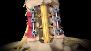
Spine Surgery encompasses a range of advanced surgical procedures designed to relieve chronic back or neck pain, decompress pinched nerves, repair herniated discs, and restore structural stability to the spinal column. From motion-preserving Artificial Disc Replacement (ADR) and microdiscectomy to complex spinal fusions, scoliosis correction, and Minimally Invasive Spine Surgery (MISS), modern spine interventions aim to restore mobility and significantly improve quality of life.

World-Class InfrastructureNABH/JCI accredited centers with global standards
Up to 80% SavingsDramatically lower cost than USA, UK, or Australia
Expert SpecialistsInternationally trained & board-certified specialists
Priority CareImmediate scheduler booking with zero waiting list

Specialist Doctor in India
500+ Top Specialists 20+ JCI & NABH Accredited Hospitals
SPECIALIZED CONDITIONS & PROCEDURES

Explore Spine Surgery Sub Categories

Explore dedicated sub-specialties with expert doctors, accredited hospital centers, and tailored treatment plans in India.

31 Sub Specialties Get Treatment Opinion
Cervical Laminectomy
Spine Surgery

Cervical Laminectomy

Cervical Laminectomy is a spinal surgery that removes part of the lamina, the bony covering at the back of the cervical vertebrae, to create additional space for the spinal cord and nerves. It may be recommended for cervical spinal stenosis, spinal cord compression, or degenerative changes causing neurological symptoms. The surgical approach depends on the location and severity of compression, spinal alignment, and stability. Recovery may include pain management, activity guidance, and rehabilitation.

Cervical Laminoplasty
Spine Surgery

Cervical Laminoplasty

Cervical Laminoplasty is a procedure that enlarges the spinal canal in the neck by reshaping and repositioning portions of the vertebral lamina. It is commonly considered for selected patients with cervical spinal stenosis and spinal cord compression, particularly when multiple levels are affected. The procedure aims to provide more space for the spinal cord while preserving more natural spinal movement than some fusion procedures. Patient selection depends on spinal alignment, stability, symptoms, and imaging findings.

Anterior Cervical Discectomy (ACD)
Spine Surgery

Anterior Cervical Discectomy (ACD)

Anterior Cervical Discectomy (ACD) is a procedure performed through the front of the neck to remove a damaged or herniated cervical disc that is pressing on a nerve root or the spinal cord. It may be considered for selected patients with persistent nerve pain, numbness, weakness, or spinal cord compression. Removing the problematic disc material helps relieve pressure on neural structures. Depending on the clinical situation, the procedure may be combined with spinal fusion or artificial disc replacement.

Anterior Cervical Discectomy and Fusion (ACDF)
Spine Surgery

Anterior Cervical Discectomy and Fusion (ACDF)

Anterior Cervical Discectomy and Fusion (ACDF) is a procedure that removes a damaged cervical disc through the front of the neck and joins the adjacent vertebrae using a bone graft or fusion material, often with an implant for support. It may be recommended for cervical disc herniation, nerve compression, or degenerative changes causing persistent symptoms. Fusion helps stabilize the treated spinal level while healing occurs. Postoperative care may include activity restrictions, follow-up imaging, and rehabilitation as advised by the surgeon.

Cervical Artificial Disc Replacement
Spine Surgery

Cervical Artificial Disc Replacement

Cervical Artificial Disc Replacement is a surgical procedure in which a damaged cervical disc is removed and replaced with an artificial implant designed to preserve motion at the treated level. It may be considered for selected patients with symptomatic cervical disc disease or nerve compression when clinical and anatomical criteria are met. Unlike fusion, the procedure aims to maintain movement between the adjacent vertebrae. Suitability depends on bone quality, spinal stability, facet joint condition, and the extent of degeneration.

Multilevel Cervical Disc Replacement
Spine Surgery

Multilevel Cervical Disc Replacement

Multilevel Cervical Disc Replacement involves replacing damaged discs at two or more levels of the cervical spine with artificial disc implants. It may be considered for carefully selected patients with symptomatic disc degeneration or nerve compression affecting multiple cervical levels. The procedure aims to relieve neural pressure while preserving movement at treated levels. Detailed assessment of spinal alignment, bone quality, joint health, and the distribution of disc disease is essential before surgery. Follow-up care monitors implant function and recovery.

Thoracic Discectomy
Spine Surgery

Thoracic Discectomy

Thoracic Discectomy is a procedure to remove herniated or damaged disc material from the thoracic spine, located in the middle portion of the back. A thoracic disc may compress the spinal cord or nearby nerves and cause pain, sensory changes, weakness, or walking difficulties. Surgery may be considered when symptoms, neurological findings, and imaging indicate significant compression. The surgical approach depends on the location and size of the disc herniation and its relationship to the spinal cord.

Thoracic Laminectomy
Spine Surgery

Thoracic Laminectomy

Thoracic Laminectomy removes a portion of the lamina from one or more thoracic vertebrae to create additional space for the spinal cord. It may be used to address spinal canal narrowing, selected spinal tumors, or other conditions causing thoracic cord compression. The surgical plan depends on the cause and extent of compression, spinal stability, and the patients neurological condition. In some cases, additional stabilization may be required. Postoperative monitoring and rehabilitation support recovery and functional improvement.

Thoracic Foraminotomy
Spine Surgery

Thoracic Foraminotomy

Thoracic Foraminotomy enlarges the opening through which a thoracic spinal nerve exits the spinal canal. The procedure may be considered when bone spurs, degenerative changes, or other structures narrow this opening and compress a nerve. By removing selected obstructing tissue, the surgeon aims to reduce nerve pressure and related symptoms. Preoperative evaluation identifies the affected level and cause of compression. Recovery instructions and rehabilitation depend on the extent of surgery and the patients overall spinal condition.

Thoracic Decompression Surgery
Spine Surgery

Thoracic Decompression Surgery

Thoracic Decompression Surgery aims to relieve pressure on the spinal cord or nerve roots in the middle portion of the spine. Compression may arise from disc herniation, spinal stenosis, abnormal bone growth, or other structural conditions. Depending on the cause, surgery may involve removing disc material, bone, or other tissue that restricts the neural structures. Treatment planning considers spinal alignment, neurological symptoms, and stability. Some patients may require additional fusion or stabilization to support the spine.

Thoracic Fusion
Spine Surgery

Thoracic Fusion

Thoracic Fusion is a surgical procedure that joins two or more vertebrae in the middle portion of the spine to provide stability. It may be performed for selected spinal deformities, fractures, instability, tumors, or degenerative conditions requiring stabilization. Bone graft material and instrumentation such as screws and rods may be used to support the fusion process. The technique depends on the underlying diagnosis and the number of spinal levels involved. Recovery includes monitoring, activity guidance, and follow-up to assess bone healing.

Posterior Thoracic Fusion
Spine Surgery

Posterior Thoracic Fusion

Posterior Thoracic Fusion joins vertebrae in the thoracic spine through a surgical approach from the back. It may be used to stabilize spinal fractures, correct selected deformities, treat instability, or support the spine after decompression or tumor surgery. Screws, rods, and bone graft material may be used to maintain alignment while the vertebrae fuse. Surgical planning considers the affected levels, bone quality, spinal cord safety, and overall health. Regular follow-up helps assess alignment, implant integrity, and fusion progress.

Lumbar Discectomy
Spine Surgery

Lumbar Discectomy

Lumbar Discectomy removes herniated material from a disc in the lower back when it compresses a nearby nerve root. It may be considered for persistent sciatica, radiating leg pain, or neurological symptoms that have not improved adequately with appropriate non-surgical treatment. The procedure aims to relieve nerve pressure while preserving as much healthy disc tissue as possible. The surgical technique depends on the size and location of the herniation. Recovery guidance may include gradual activity, exercises, and follow-up assessment.

Microdiscectomy
Spine Surgery

Microdiscectomy

Microdiscectomy is a minimally invasive or limited-incision procedure used to remove the portion of a herniated spinal disc that compresses a nerve. It is commonly performed in the lumbar spine to treat selected cases of sciatica and radiating leg pain. Magnification and specialized instruments help the surgeon work around delicate neural structures. The procedure aims to relieve nerve irritation while minimizing disruption to surrounding tissues. Recovery varies, and patients receive guidance on movement, lifting, rehabilitation, and warning signs that require medical review.

Endoscopic Lumbar Discectomy
Spine Surgery

Endoscopic Lumbar Discectomy

Endoscopic Lumbar Discectomy uses a small endoscope and specialized instruments to remove herniated disc material from the lower back. It may be suitable for selected lumbar disc herniations causing nerve compression and persistent leg pain. The endoscopic approach allows the surgeon to visualize the treatment area through a small access channel, potentially reducing soft-tissue disruption in appropriate cases. Suitability depends on disc location, spinal anatomy, and clinical findings. Postoperative instructions and rehabilitation are individualized to support recovery.

Lumbar Laminectomy
Spine Surgery

Lumbar Laminectomy

Lumbar Laminectomy removes part or all of the lamina from one or more lower-back vertebrae to increase space in the spinal canal. It may be recommended for lumbar spinal stenosis that compresses nerve structures and causes leg pain, numbness, weakness, or walking limitations. The operation is planned according to the location and severity of narrowing and the stability of the spine. Fusion may be considered when clinically indicated. Recovery commonly includes gradual mobilization, exercise guidance, and follow-up care.

Lumbar Decompression
Spine Surgery

Lumbar Decompression

Lumbar Decompression Surgery relieves pressure on nerves in the lower back caused by spinal stenosis, disc herniation, bone spurs, or other structural changes. Depending on the diagnosis, treatment may involve removing a small amount of bone, thickened ligament, or disc material. The goal is to reduce nerve compression and improve related pain, sensation, strength, or walking ability. Surgical planning takes into account spinal stability, the number of affected levels, and the patients overall health. Rehabilitation is tailored to individual needs.

Endoscopic Discectomy
Spine Surgery

Endoscopic Discectomy

Endoscopic Discectomy is a minimally invasive technique that uses an endoscope and specialized instruments to remove problematic disc material pressing on a spinal nerve. It may be used for selected herniated discs, most commonly in the lumbar spine, depending on the location and anatomy of the affected disc. A small access channel allows visualization of the surgical area while limiting disruption to surrounding tissues. The appropriate approach depends on clinical assessment and imaging. Recovery instructions are individualized to the procedure and patient.

Endoscopic Foraminotomy
Spine Surgery

Endoscopic Foraminotomy

Endoscopic Foraminotomy uses an endoscope and specialized instruments to enlarge a narrowed opening through which a spinal nerve exits the vertebral column. It may be considered for selected cases of foraminal stenosis caused by degenerative changes, bone spurs, or disc-related compression. The procedure aims to reduce pressure on the affected nerve while limiting disruption to nearby tissues. The spinal level, severity of narrowing, and stability of the spine influence treatment selection. Follow-up care and rehabilitation depend on the individual case.

Endoscopic Decompression
Spine Surgery

Endoscopic Decompression

Endoscopic Decompression is a minimally invasive spinal procedure that uses an endoscope and specialized instruments to relieve pressure on compressed nerves or the spinal canal. Depending on the underlying condition, the surgeon may remove selected bone, ligament, or disc material. It may be considered for appropriately selected cases of spinal stenosis or nerve compression. The technique and expected recovery depend on the affected spinal region, anatomy, and extent of disease. Clinical assessment and imaging help determine whether an endoscopic approach is appropriate.

Full-Endoscopic Spine Surgery
Spine Surgery

Full-Endoscopic Spine Surgery

Full-Endoscopic Spine Surgery uses an endoscope with a camera and specialized instruments to perform selected spinal procedures through a small access pathway. Depending on the diagnosis, it may be used for disc herniation, nerve compression, or certain forms of spinal stenosis. The technique can reduce muscle disruption in appropriately selected patients, although it is not suitable for every spinal condition. Procedure selection depends on anatomy, disease severity, and surgeon expertise. Recovery and activity recommendations are tailored to the operation performed.

Percutaneous Pedicle Screw Fixation
Spine Surgery

Percutaneous Pedicle Screw Fixation

Percutaneous Pedicle Screw Fixation stabilizes the spine by inserting screws into the vertebral pedicles and connecting them with rods through small skin incisions. It may be used for selected spinal fractures, instability, deformity correction, or as part of a fusion procedure. Image guidance helps the surgical team place implants accurately. Compared with traditional open approaches, this technique may reduce muscle disruption in suitable cases. The need for fusion, postoperative restrictions, and rehabilitation depends on the underlying spinal condition.

Percutaneous Fusion
Spine Surgery

Percutaneous Fusion

Percutaneous Spinal Fusion uses small incisions and specialized instruments to stabilize selected spinal segments and support fusion between vertebrae. Instrumentation such as screws and connecting rods may be inserted through the skin, sometimes alongside other procedures that prepare the bone for fusion. It may be considered for selected cases of instability, fractures, or degenerative spinal conditions. The exact technique depends on the diagnosis, spinal levels, and bone quality. Follow-up care monitors healing, implant position, and functional recovery.

Minimally Invasive TLIF
Spine Surgery

Minimally Invasive TLIF

Minimally Invasive Transforaminal Lumbar Interbody Fusion (TLIF) is a procedure that removes a damaged lumbar disc through a posterior or posterolateral approach and places bone graft material or an interbody implant between the vertebrae. Screws and rods may be used to stabilize the treated level while fusion develops. It may be recommended for selected cases of spinal instability, degenerative disc disease, or certain forms of spondylolisthesis. Treatment planning and recovery depend on spinal anatomy, bone health, and the patients clinical needs.

Minimally Invasive Laminectomy
Spine Surgery

Minimally Invasive Laminectomy

Minimally Invasive Laminectomy removes selected portions of bone and other obstructing tissue through a smaller surgical opening to relieve pressure on spinal nerves or the spinal cord. It may be used for suitable cases of spinal stenosis or nerve compression. Specialized instruments and tissue-preserving techniques can reduce disruption to surrounding muscles in selected patients. The best approach depends on the affected spinal level, degree of narrowing, and stability. Postoperative care includes progressive mobilization and individualized rehabilitation.

Minimally Invasive Microdiscectomy
Spine Surgery

Minimally Invasive Microdiscectomy

Minimally Invasive Microdiscectomy removes the portion of a herniated spinal disc that is compressing a nerve through a small incision using magnification and specialized instruments. It is commonly considered for selected lumbar disc herniations associated with persistent sciatica or neurological symptoms. The procedure aims to relieve nerve pressure while limiting damage to nearby muscles and soft tissues. Suitability depends on the location of the herniation and the patients symptoms. Recovery guidance includes activity progression, lifting precautions, and follow-up care.

Scoliosis Surgery
Spine Surgery

Scoliosis Surgery

Scoliosis Surgery is performed to correct or stabilize an abnormal sideways curvature of the spine in selected patients. Surgery may be considered when a curve progresses substantially, causes significant imbalance or symptoms, or presents other clinically important concerns despite appropriate monitoring or conservative treatment. Depending on the curve and patient characteristics, treatment may involve spinal instrumentation and fusion, with the aim of improving alignment and preventing further progression. Planning considers skeletal maturity, curve pattern, neurological status, and overall health.

Intradural Tumor Resection
Spine Surgery

Intradural Tumor Resection

Intradural Tumor Resection is a specialized spinal operation to remove or reduce a tumor located within the dura, the protective membrane surrounding the spinal cord and nerve roots. Such tumors may cause pain, weakness, sensory changes, or other neurological symptoms depending on their location and size. Preoperative imaging helps define the tumor and its relationship to neural structures. The surgical approach is selected to provide appropriate access while protecting spinal cord and nerve function. Further treatment and monitoring depend on the tumor diagnosis and pathology results.

Extradural Tumor Resection
Spine Surgery

Extradural Tumor Resection

Extradural Tumor Resection removes or reduces a tumor located outside the dura surrounding the spinal cord. These tumors may arise from spinal bones, surrounding tissues, or spread from another site in the body. Treatment planning depends on tumor type, location, spinal stability, neurological symptoms, and the extent of disease. Surgery may aim to relieve neural compression, obtain tissue for diagnosis, remove accessible tumor tissue, or stabilize weakened vertebrae. Additional oncology treatment and follow-up may be required according to the final diagnosis.

Spinal Cord Tumor Removal
Spine Surgery

Spinal Cord Tumor Removal

Spinal Cord Tumor Removal is a specialized procedure intended to remove or reduce a tumor affecting the spinal cord or its surrounding structures. Symptoms may include back pain, weakness, altered sensation, balance difficulties, or changes in bladder and bowel function. The surgical plan depends on tumor location, imaging findings, neurological status, and the relationship between the tumor and healthy neural tissue. The goal is to treat the tumor while preserving neurological function whenever possible. Pathology results guide decisions about additional treatment and surveillance.

Nerve Sheath Tumor Excision
Spine Surgery

Nerve Sheath Tumor Excision

Nerve Sheath Tumor Excision is a surgical procedure to remove a tumor arising from the protective tissues surrounding a peripheral nerve or spinal nerve root. Many such tumors are benign, although diagnosis requires appropriate clinical evaluation and, when indicated, tissue examination. Surgery may be recommended when a tumor causes pain, progressive neurological symptoms, significant compression, or other concerning features. The surgical approach aims to remove the lesion while protecting nerve function. Follow-up depends on pathology findings, the extent of removal, and the patients recovery.

OUR SPECIALISTS

Top Specialists in Spine Surgery

View All Doctors
Principal Director & Head – Orthopaedics & Spine Spine Surgery | Orthopaedics | Joint Replacement

Dr. (Prof.) Bhavuk Garg

Academic Qualifications & Advanced Training Dr. Bhavuk Garg has undertaken extensive medical and specialist training in India and the United States. His qualifications and fellowships include: MBBS – University College of Medical Sciences, New Delhi MS
25+ Years Experience
Max Smart Hospital (Max Saket),
★★★★★ 4.8
View Profile & Book
Director & Head - Minimal Access, Bariatric & Gl Surgery Ex Professor of Surgery, AlIMS, New Delhi.

Dr. A. K. Kriplani

MBBS, MS (General Surgery)
30 Experience
All India Institute of Medical Sciences (AIIMS),
★★★★★ 4.8
View Profile & Book
Senior Director – Orthopaedics & Joint Replacement | Spine Surgery

Dr. Harshavardhan Hegde

Academic Qualifications Dr. Harshavardhan Hegde completed his: MBBS – 1983 MS (Orthopaedics) – 1987, Baroda Medical College Following his postgraduate training, he pursued additional specialist training and fellowships in orthopaedic and spinal surgery
40+ Years Experience
Max Smart Hospital (Max Saket), Delhi
★★★★★ 4.8
View Profile & Book
Director – Medical Oncology | Cancer Specialist in Delhi NCR

Dr. Nagender Sharma

Academic Qualifications & Medical Training Dr. Nagender Sharma's medical education includes: MBBS – Kasturba Medical College, Mangalore MD in Medicine – M. R. Medical College, Gulbarga DM in Medical Oncology – Cancer Institute (WIA), Adyar, Chennai DNB
14+ Years Experience
Max Smart Hospital (Max Saket), Delhi
★★★★★ 4.8
View Profile & Book
WHERE TO GET TREATED

Accredited Hospitals for Spine Surgery

View All Hospitals

Amrita Hospital

Leading multi-specialty healthcare services in India.

Faridabad, Haryana Est:
JCINABHNABL
View Hospital Details

Apollo Athenaa Women’s Cancer Centre

Dedicated Women’s Cancer Care, Bringing Together Specialist Expertise, Advanced Treatment and Compassionate Support for Every Patient

Delhi, Delhi NCR Est: 2025
JCINABH
View Hospital Details

Apollo Hospitals

Leading multi-specialty healthcare services in India.

Gurugram, Haryana Est: 2026
NABH
View Hospital Details
Specialty Patient recovery
SUCCESS STORY

Real Recovery.
Real Hope.

"
★★★★★

Coming from Kenya to seek medical treatment in India was a daunting step. But the team at AthelCare supported us at every single moment—from visa documentation, airport pickup, hospital appointments, to recovering comfortably. The cost was extremely affordable and the surgical outcome was absolutely successful. Highly recommended!

JM
Joseph Mwangi 🇰🇪 Kenya — Patient Recovery Success Treated at Apollo Hospitals, New Delhi
FAQ

Common Questions

Get Free Consultation
How do I request a cost estimate or medical opinion?
Simply upload your diagnostic reports and medical summary using our contact form. Our specialized clinical team will review them and send you a detailed treatment proposal and pricing quote within 24-48 hours.
How long will I need to stay in India for treatment?
This varies depending on the severity of the condition and surgical requirements. Minor surgeries require 1-2 weeks of stay, while major surgeries or transplants require 4-8 weeks.
Are clinical results and facilities comparable to USA/UK?
Yes. India's top super-specialty hospitals are NABH & JCI accredited. They leverage state-of-the-art diagnostic equipment, robotic-assisted surgical platforms, and are staffed by highly qualified, fellowship-trained experts.
YOUR HEALTH IS OUR PRIORITY

Take the first step toward
a healthier life.